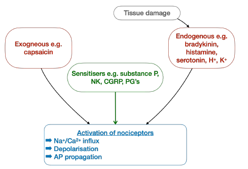
The syllabus asks for knowledge of "afferent nociceptive pathways, dorsal horn, peripheral and central mechanisms, neuromodulatory systems, supraspinal mechanisms, visceral pain, neuropathic pain, influence of therapy on nociceptive mechanisms".
This carries through to the Final syllabus, which lists the "anatomy of ascending and descending pain pathways and their relevance to the pharmacological management of pain".
The March 2017 CRQ on persistent post-surgical pain included a section on the pathophysiological changes that occur at spinal cord level during the transition from acute to persistent pain; i.e. wind-up and central sensitisation, covered below.
When answering a question on pain pathways, a useful structure is to follow the journey of the stimulus:
Transduction: conversion of a noxious stimulus into an action potential at the nociceptor
Transmission: conduction to the dorsal horn and up to the thalamus
Modulation: amplification or inhibition of the signal, mainly at the dorsal horn
Perception: conscious awareness and interpretation of pain in the cortex
Each stage is a target for analgesic therapy
Nociceptors
Nociceptors are free, typically unmyelinated nerve endings that are abundant in skin and musculoskeletal tissue
They may be activated by noxious stimuli which are:
Exogenous e.g. capsaicin
Endogenous i.e. mediators released by tissue damage: bradykinin, histamine, serotonin, ACh, H+ and K+
Activation causes Na+/Ca2+ influx, depolarisation and initiation of an action potential
Activation and sensitisation of nociceptors
Peripheral sensitisation
Some mediators sensitise nociceptors by reducing their threshold for activation:
Substance P, prostaglandins, leukotrienes, neurokinin A and CGRP
Neighbouring nociceptors are sensitised, producing a zone of primary hyperalgesia around the injury
Low-intensity stimuli may now produce pain, with increased sensitivity to thermal stimuli
Aβ
Aδ
C
Myelination
Myelinated
Thinly myelinated
Unmyelinated
Stimulus
Non-noxious Touch/pressure
Unimodal noxious Mechanical/thermal
Polymodal noxious Mechanical, thermal, chemical
Conduction (m/s)
80-120
15-30
0.5-2
Quality of pain
N/A
'First' pain: sharp, well localised, short lasting
'Second' pain: dull, burning, poorly localised
Dorsal horn termination
Lamina III-V
Laminae I and V
Laminae I and II (substantia gelatinosa)
Neurotransmitter
Glutamate
Glutamate
Glutamate Substance P
First order neurones
The action potential travels along Aδ and C fibres, whose cell bodies lie in the dorsal root ganglion, to the dorsal horn of the spinal cord
At the pre-synaptic terminal, Ca2+ influx causes release of excitatory neurotransmitters:
Glutamate: acting at AMPA, kainate and NMDA receptors
Aspartate: acting at NMDA receptors
Substance P: acting at NK-1 receptors
The dorsal horn
First order neurones synapse with second order neurones in Rexed's laminae of the dorsal horn
Two types of second order neurone exist:
Nociceptive-specific neurones: respond only to noxious stimuli; found in the superficial laminae
Wide dynamic range neurones: respond to both noxious and non-noxious stimuli; found in the deeper laminae (IV and V)
Central sensitisation
Repeated C fibre input removes the Mg2+ block of the NMDA receptor, increasing the sensitivity of dorsal horn neurones
This underlies the medium- and long-term changes seen in chronic pain states:
Wind-up: a progressive increase in dorsal horn neurone response to repeated stimuli, greater than would be expected from a linear stimulus-response relationship
Central sensitisation: low-intensity stimuli produce pain, with a zone of secondary hyperalgesia in uninjured tissue
NB unlike peripheral sensitisation, there is no increased response to thermal stimuli
Long-term potentiation and altered gene expression
Ascending tracts
Most second order neurones decussate and ascend in the anterolateral system
Tracts are somatotopically organised; fibres from caudal segments lie laterally and those from rostral segments medially
Spinothalamic tract
Lateral (pain and temperature) and anterior (crude touch) divisions
Fast, discriminative fibres project directly to the ventral posterior nucleus of the thalamus
Slow, affective fibres synapse in the reticular formation and intralaminar thalamic nuclei, then project to the hypothalamus, limbic system and autonomic centres
Spinoreticular tract: terminates in the reticular formation; arousal, motor and autonomic reflexes
Spinomesencephalic tract: terminates in the PAG; integration of pain responses and autonomic reflexes
For comparison, touch, vibration and proprioception (Aβ) ascend ipsilaterally in the dorsal columns before decussating in the medulla to form the medial lemniscus
Withdrawal reflex
In parallel, a protective polysynaptic withdrawal reflex occurs at spinal cord level, before the stimulus is perceived
Inhibitory neurotransmitters
GABA
Endogenous opioids e.g. enkephalins acting at MOP receptors
Opioid receptors are pre-synaptic Gi protein-coupled receptors
Following tissue damage, they are synthesised in the dorsal root ganglion and transported peripherally along the sensory nerve, forming a peripheral mechanism of analgesia
Segmental modulation
Transmission at the dorsal horn is influenced by other afferent inputs, as described by the gate control theory
Descending inhibitory pathways
Act as negative feedback, opposing the positive feedback of peripheral and central sensitisation
Descend in the dorsolateral funiculus
Periaqueductal grey (PAG)
Midbrain area projecting to the nucleus raphe magnus (NRM) in the medulla, which in turn projects to the dorsal horn
Serotonergic (5-HT)
Locus coeruleus (LC)
Brainstem nucleus projecting to the dorsal horn
Noradrenergic (α2-adrenoceptors)
Both pathways activate inhibitory enkephalinergic interneurones in the substantia gelatinosa, reducing onward transmission
Second order neurones synapse with third order neurones in the ventral posterior and intralaminar nuclei of the thalamus
Third order neurones project to:
Primary somatosensory cortex (postcentral gyrus of the parietal lobe): location and intensity of pain
Anterior cingulate and frontal cortex: emotional aspects of pain
Limbic system, hypothalamus and autonomic centres: affect, memory and autonomic responses
Perception is therefore shaped by mood, expectation and prior experience, which is the basis of the biopsychosocial model of pain
Site
Analgesic interventions
Periphery (transduction)
NSAIDs Local anaesthetics Peripheral opioid receptors